Welcome to Xuzhou Ruihua Electronic Technology Development Co., Ltd.!
Provide overall solutions for medical devices
More than 20 years of production experience, hot sale nationwide

National consultation hotline:
Product Center
Details description
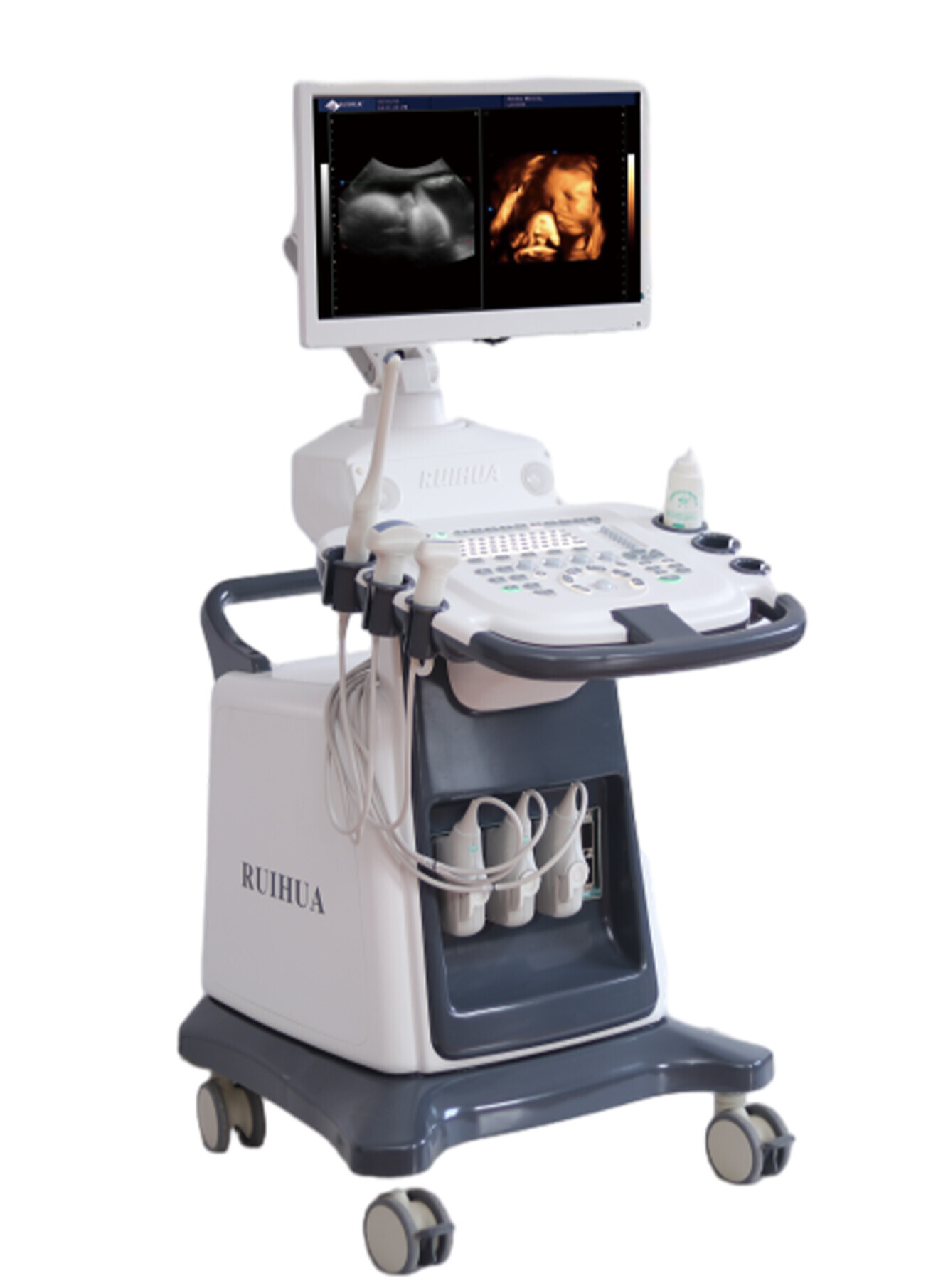
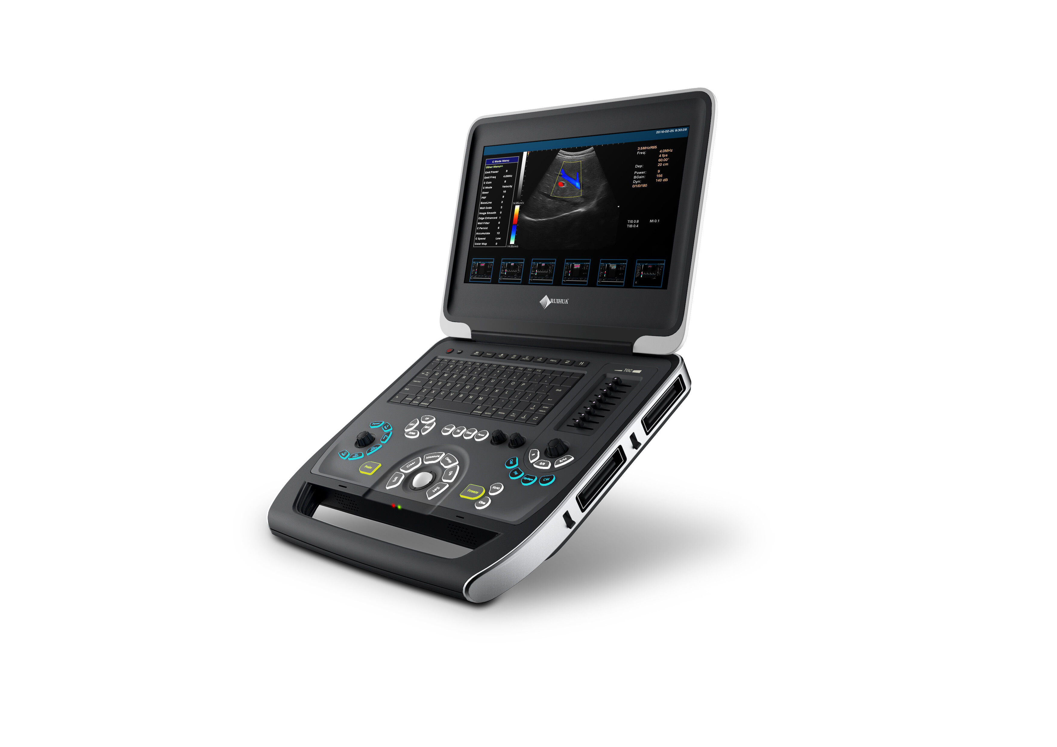
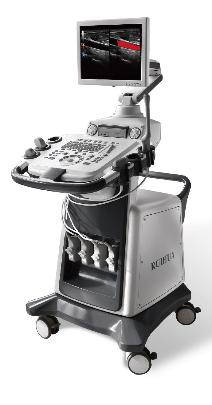
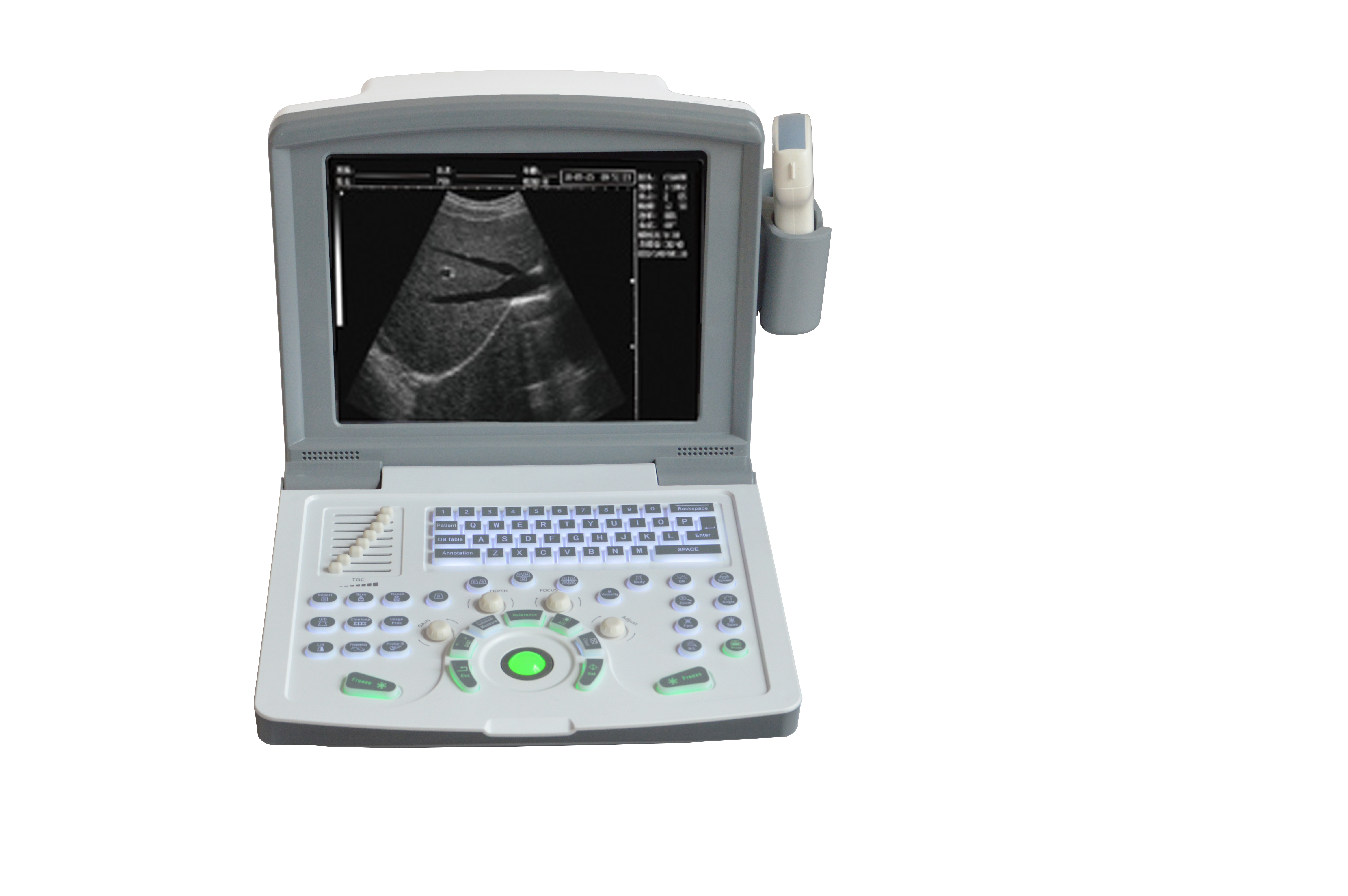
Application range : It is suitable for diagnosis and analysis of the abdomen (liver, gallbladder, kidney, pancreas, spleen), obstetrics and gynecology, heart, thyroid, small organs, urology, superficial and orthopedic surgery. It can perform power diagnosis on large blood vessels such as liver, gallbladder, spleen, kidney and carotid artery.
Imaging unit Digital 2D grayscale imaging unit
Digital Color Doppler Unit
Digital Spectral Doppler Display and Analysis Unit
Digital power blood flow imaging unit
Color Doppler operating frequency Color and Doppler operating frequency can be digitally displayed and adjusted independently
Beam processing: fully digital beam former, continuous dynamic focusing, dynamic variable aperture, dynamic beam apodization, dynamic filtering;
Digital beam enhancer, multiple beam synthesis, inverted pulse harmonic imaging, Doppler imaging (including color, energy, directional energy Doppler mode), spectral Doppler imaging (including pulse Doppler, continuous wave Doppler), speckle noise suppression technology, speckle softening technology
Extended pulse Extended pulse imaging technology improves image penetration and increases the ability to examine difficult patients in the far field
Probe array element number ≥ 128
Image magnification : real-time partial magnification, shiftable, 6 times magnification, 16 levels adjustable
Image pre-processing (total gain, acoustic power, gray value, dynamic range);
Post-processing (edge enhancement, frame averaging, line averaging, gamma correction, contrast, brightness);
Correlation processing, logarithmic compression, TGC control, interpolation, black and white grayscale flip, left and right flip, up and down flip, frame correlation, smoothing, black and white grayscale conversion, grayscale suppression
Image pseudo color function has image pseudo color function
Movie playback image retrieval, movie loop playback: ≥ 1536 frames, playback time ≥ 75 seconds
Chinese and English interface display: hospital name, department, patient ID , name, date, time, probe number, frequency, gain, depth, focus position, magnification, puncture guide line, text annotations, etc.
Basic Measurements
Calculation function: B- mode basic measurements: distance, angle, perimeter and area, volume, stenosis rate, histogram, cross-section
M mode basic measurement: heart rate, time, distance, speed
Doppler measurements: time, heart rate, velocity, acceleration
Routine general measurement: distance, area, perimeter, volume, ratio, angle, histogram, cross-section, residual urine volume, stenosis rate, heart rate, time, slope speed
Heart measurements and calculations Time, slope, heart rate, left ventricle measurement, left ventricular function, wall thickness assessment, mitral valve measurement, aorta measurement, body surface area calculation, heart measurement report
The system presets of the inspection mode provide real-time, non-preset 2D, color Doppler and spectral Doppler modes. One-touch automatic image optimization adjustment
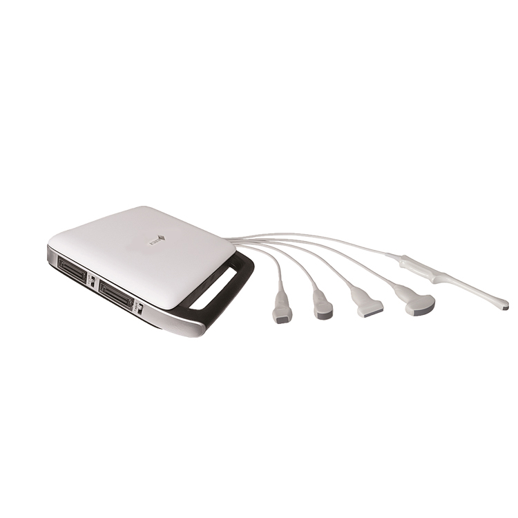
The probe is connected to 4 interfaces, all activated and automatically identified
Probe receiving dynamic range ≥ 150dB ( 40dB ~ 150dB visible, adjustable)
Interface language : Chinese / English interface, switchable, Chinese / English input possible
Products Recommended
Online Consultation

